
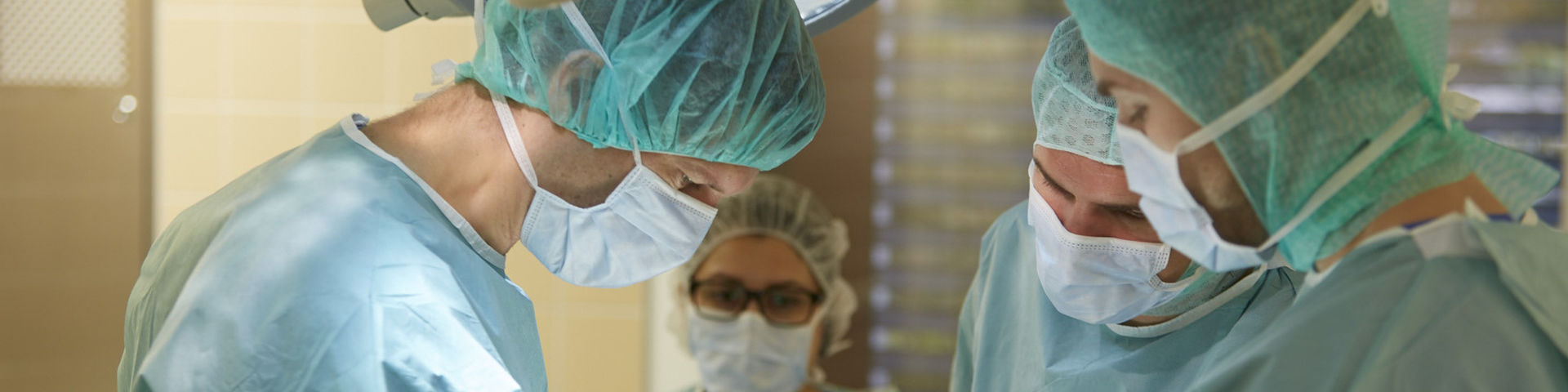
Osteoarthritis | Operative Treatment
Joints must work reliably for many decades day after day, and in some cases withstand extreme stress. A healthy cartilage system is necessary in order for the joints to function optimally. Cartilage not only ensures the smooth movement of the joint but also acts as a shock absorber enduring loads and stress up to seven times our body weight. If in case of a knee arthrosis, conservative treatment options have been exhausted or a significant deterioration of the condition is to be expected, surgery should be considered.
 Video: Kniegelenk Arthrose Knorpelschaden OP
What is cartilage damage?
Damage to the joint cartilage can be age-related, as a result of excessive loading (sports, work, overweight), lack of exercise, torn ACL, torn meniscus or sports accidents. Treatment is often difficult because the cartilage is not supplied with blood and tissue, and therefore lacks the ability to regenerate itself.
The loss of cartilage is irreversible, meaning cartilage damage does not heal. More severe cartilage defects may lead to osteoarthritis (significant joint damage) if left untreated. More extensive cartilage damage requires therefore treating the cartilage surgically in order to prevent osteoarthritis. An option is arthroscopic surgery, i.e. minimally invasive surgical procedures. During arthroscopy, surgery is performed using a mini camera via two small incisions 2-3 mm each. What is the best method to treat cartilage damage depends on the age and on the patient's expectations, e.g. activity level. However, it depends primarily on the extent of the cartilage damage. Cartilage damages is measured in grades I to IV .
- Grade I the cartilage has softened
- Grade II the cartilage has a rough surface, e.g. has minor tears
- Grade III lesions have deep crevices
- Grade IV the cartilage is graded down to expose the underlying (subchondral) bone
Arthroscopic procedures for the treatment of cartilage damage
Essentially the following arthroscopic surgical procedures are available to treat articular cartilage damages
- Cartilage smoothing at Grade II - III cartilage damage
- Microfracture / abrasion arthroplasty for Grade IV cartilage damage
- Osteochondral transplantation (OCT) for Grade IV cartilage damage
- Autologous cartilage cell transplantation (ACT) for Grade IV cartilage damage
Cartilage smoothing
During cartilage smoothing, only the roughness of the joint surface is carefully smoothed arthroscopically. The aim is a maximum cartilage preservation to mitigate the progression of cartilage damage. Moreover, within the scope of the procedure, the cartilaginous abrasive particles are rinsed out of the joint in order to reduce irritation of the joint.
Microfracture / abrasion arthroplasty
In the process known as bioprosthesis, a profound cartilage defect (grade IV) is filled with endogenous tissue. In medical terms, this is called microfracturing (alternatively, abrasion arthroplasty, pridie drilling or chondral picks). Here the bare bone is "injured" so that bone bleeding takes place. The process is similar to a skin injury, where a scar forms during the healing process: a scar of fibrocartilage forms in the joint due to the stem cells exiting from the bone marrow. This cartilaginous repair tissue fills and seals the defect cartilage (bioprosthesis). In testing, it is essential to ascertain whether the concentration of stem cells increase and thus an improved healing of the defect can be achieved through the additional covering of the defect with an exogenous 3-dimensional fleece (non-woven), the so-called chondrotissue.
Arthroscopic and microscopic examinations show a convincing defect fill for smaller and mid-size defects, in particular if the opposite joint surface is for the most part intact.
Cartilage – bone transplantation / osteochondral transplantation (OCT)
In osteochondral transplantations also called OCT or OATS, osteochondral cylinders are taken from a less weight-bearing articular surface and transplanted into the drilled defect area. Thus, a large part of a highly strained defect surface can be filled with high-quality hyaline cartilage. The remaining gaps between the cylinders are covered with scar tissue. The results are good; however, in this procedure healthy cartilage surfaces are destroyed. This may cause discomfort to the cylinder outlet. To keep these symptoms to a minimum, the APR should be performed in cartilage defects of max. 2 – 3 qcm.
Endogenous cartilage cell transplantation / autologous chondrocyte transplantation (ACT)
In a chondrocyte transplantation, cartilage cells are extracted arthroscopically from the patient's healthy articular cartilage that is located in a non-load-bearing area and then propagated in a special laboratory. After about 3 - 4 weeks, a sufficient amount of cell material is available. With the help of the body biodegradable carrier tissue, shortly before the transplantation this material is formed to accurately fit the defect. During a second surgery, the existing cartilage defect is first cleaned from the worn cartilage. The preformed graft is then cut to the desired size, and with the aid of anchoring sutures inserted precisely into the defect. The cartilage cells have a chance to “heal into” the defect.
Although, a chondrocyte transplantation assures a complete healing of cartilage damage, at this time only patients with locally circumscribed cartilage damage can be treated successfully. Patients must be carefully selected to insure treatment according to the damage. The procedure can currently not be applied in patients with advanced joint degeneration, e.g. osteoarthritis. However, further development of this innovative treatment method is anticipated in the future, which would allow using the method within a broad spectrum of anterior cartilage damages.
Post surgical treatment arthroscopic cartilage procedure
Independent of which procedure is being used, as a rule, the joint should be mobilized at an early stage during post-treatment, i.e. rehabilitation. However, depending on the duration of surgical procedures, rehabilitation measures might differ and the joint should not be strained in the early stage after the surgery. In most cases, the patient will have to walk on crutches. After smoothing of the joint surface or during implantation of a TRUFIT cylinder, the joint is fully functional again after 1-2 weeks. After cartilage stimulating procedures and of cartilage cell transplantation, the joint must principally remain free from strain for several weeks, usually this means a period of 6-8 weeks.
As a part of the rehabilitation, targeted physical therapy includes injections with hyaluronic acid and dietary supplements such as Glucosamine and chondroitin to support the regeneration of the joint cartilage.
What is the best procedure for an operative anterior cartilage treatment?
Which treatment is best for cartilage damage, what the risks are, and what result can be expected, must be discussed with the consulting orthopedist after a comprehensive preliminary examination. The specialist team of the KLINIK am RING in Cologne has extensive experience in the treatment of osteoarthritis and highly values individual consultation and optimal individualized treatment strategies.